down syndrome baby at 14 weeks ultrasound
An ultrasound scan could save many mothers the decision over whether to have an amniocentesis and risk losing a baby. Most doctors do an ultrasound early in the second trimester between 16 and 20 weeks.
Pin On Ob
Im 31 and very worried.

. This week we had two scheduled ultrasounds. This doesnt mean the baby has Down syndrome but. Nearly two-thirds of 15-22-week-old fetuses with Downs syndrome lack a nasal.
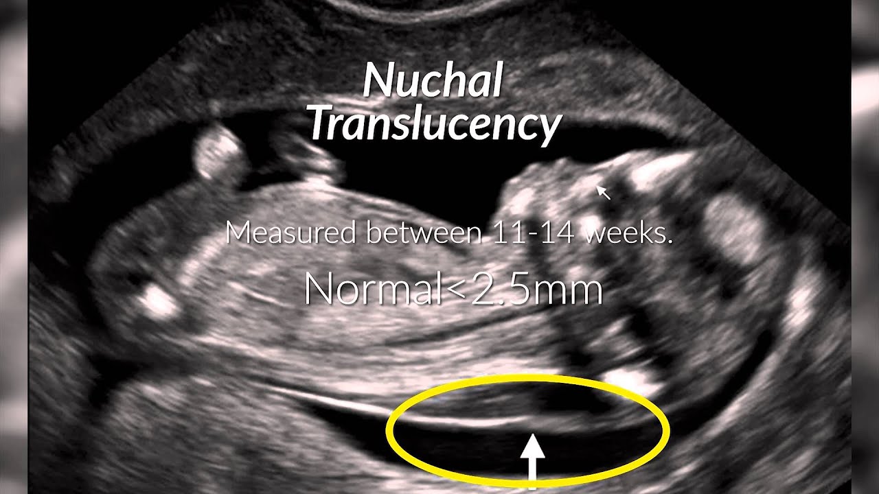
Pregnant woman should have an ultrasound done at 11 14 weeks one scan during second trimester. If the nuchal translucency test indicates that your baby may have a health condition you can decide whether to have a diagnostic test to find out for certain. Ultrasound PT and NBL determination was performed in 504 normal fetuses and 17 fetuses with Downs syndrome DS.
NT focuses on a small clear space at the back of a growing babys neck called the nuchal fold. Typically a routine scan to check on development is done during this time frame anyway. A nuchal translucency screening or NT screening is a specialized routine ultrasound performed at the end of the first trimester of pregnancy.
However ultrasound is often used as a screening test for Down syndrome and other chromosome abnormalities. Soft markers for Down syndrome are found on ultrasound scans done during the second trimester of pregnancy. A Detailed Anomaly Scan done at 20 weeks can only detect 50 of Down Syndrome cases.
For the first 14 weeks of pregnancy the evidence supports the use of first trimester ultrasound tests in combination with two serum blood markers - especially pregnancy-associated plasma protein A PAPP-A and free beta human chorionic gonadotrophin ßhCG - and maternal age for Downs syndrome screening. An ultrasound can detect birth defects like downs syndrome patau syndrome triploidy edwards syndrome turners syndrome. The NT scan is an ultrasound done in the first trimester to determine your babys risk of having Down syndrome and some other chromosomal abnormalities.
First trimester screening is a prenatal test that offers early information about a babys risk of certain chromosomal conditions Down syndrome trisomy 21 and extra sequences of chromosome 18 trisomy 18. Down syndrome in ultrasound. It helps doctors determine if a baby is statistically more likely to have a chromosomal abnormality.
The first was the nuchal translucency scan which determines whether or not the baby appears to have Down Syndrome. And d position negative in a trisomy-18 fetus at 23 5. Ultrasound and blood test and the fluid level was 27 and the blood test showed I have a 1 in 200 chance of having a baby with Down syndrome.
The ultrasound examination cannot diagnose a fetus with Down syndrome with certainty. Best of luck to you. The doctor is.
Two-dimensional ultrasound images of fetal profile FP line at. One or more abnormalities were found in 31 fetuses 33 including two of 11 fetuses seen before 14 weeks 17 of 68 fetuses seen between 14-24 weeks and 12 of 15 fetuses seen after 24 weeks. Fortunately everything else on ultrasound measured and looked perfectly.
A woman must be 11 weeks 2 days pregnant to 14 weeks one day pregnant to have this scan for the best results. 14 Week Ultrasound GENDER REVEAL. Keep in mind that an ultrasound done around the 20 th week often has signs of soft markers or around 1 in 30.
I met with a genetic counselor and got blood test done that will determine with 99 accuracy if my baby does have DS. Absent or shortened nasal bone this marker has a stronger link with Down Syndrome than most others. First of all the timing of this particular scan is very important.
I have been a nervous wreck since I was informed that my baby had a small white bright spot on her heart which is a soft marker for Down syndrome. Ashley and I were not really concerned regarding this because Down Syndrome. Birth defects have been major problem worldwide ultrasound has played an important role in detection of these.
Measurements were made from mid-sagittal 2D images acquired using a standardized. Its usually done along with a blood test. At the moment there still isnt a completely safe test that will tell you that your baby definitely does or doesnt have Downs syndrome but the NHS offers everyone combined first trimester screening which is a test performed at around 12 weeks using a combination of ultrasound scan findings and a basic.
To perform a multicenter prospective study of ultrasound prenasal thickness PT and nasal bone length NBL measurement at 11-14 weeks gestation. One soft marker that might have shown up on the first-trimester NT screening which is always performed between weeks 10 and 13 is nuchal-fold thickening where the area at the back of a babys neck accumulates fluid causing it to appear. B position zero in a fetus with Down syndrome at 21 3 weeks.
This causes a wide range of both physical disability and learning difficulties. A position zero in a euploid fetus at 24 6 weeks gestation. I am so excited that I get to post this.
During this time the babys length from crown to rump is between 45 mm and 84 mm. First Trimester Screening using bloods and Nuchal Translucency measurement done between 10-14 weeks can detect 94 of cases and Non-invasive Prenatal Testing NIPT from 9 weeks can detect 99 of Down Syndrome cases. Certain findings sometimes called soft markers on ultrasound may make your doctor more suspicious that your baby may have Down syndrome.
We did have an anatomy scan 20 weeks as we waiting for results and no markers were found. C position positive in a fetus with Down syndrome at 28 2 weeks. Prenatal ultrasound findings were reviewed in 94 consecutive fetuses with proved Down syndrome trisomy 21 during a 6-year period at a single institution.

Fetal Medicine Foundation Nasal Bone Medicine Fetal Sonography

Pin On 4d Baby Scan

14 Weeks Faces I Had My 14 Week Ultrasound Today And Found Out That I Am Having Identical Twin Twins Ultrasound Baby Ultrasound Pictures Ultrasound Pictures

20 Week Ultrasound Our Baby Girl Is Having A Baby Boy Having A Baby Boy Ultrasound Boy Or Girl Having A Baby

Pin On Pregnancy Hacks

Pin On Ultrasound

Fetal Ultrasound Image Composite Created During Exams To Screen For Spina Bifida Katherine Fornell Ultrasound Fetal Ultrasound Pictures

3d 3rd Trimester Ob Ultrasound Images 3d 4d Ultrasound Performed In The Early Third Trimester Usually Provide Images That Look More Like A Baby And Less Li

Amniotic Band Syndrome Abs Ultrasound Obstetric Ultrasound Ultrasound Sonography

Nuchal Translucency Certification Needed For High Risk Ob Ultrasound Technician Ultrasound Sonography Diagnostic Medical Sonography

Another Ultrasound Turner Syndrome Awareness Turner Syndrome Will Turner
Pin On Pregnancy Parenting

Pin On Prenatal Screening

Pin On Baby Syron

Pin On Our Baby

Pin On Stuff

Pin On Bebekler

Nuchal Translucency Scan 11 Weeks 14 Weeks Nuchal Translucency Scan Prenatal Screening

Pin On Ultrasound